
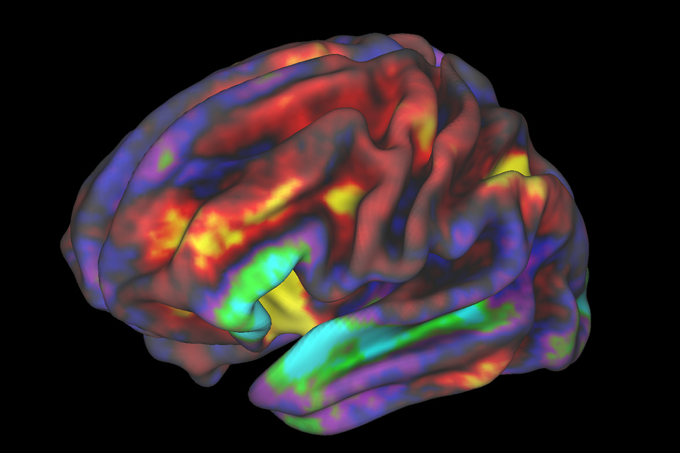
Perusing through recent discoveries in cognitive science, it’s nearly impossible not to stumble upon 3D images of the brain with certain areas lit up in varying shades of blues, reds, and yellows. These images would not be possible without neuroimaging, or techniques that allow scientists to take a literal picture what’s happening inside someone’s head. These techniques were first introduced to the scientific community in the late 1980s to early 1990s, and the number of articles published related to neuroimaging has increased nearly exponentially since then (1). Neuroimaging has transformed fields as diverse as biochemistry, marketing, and psychology, allowing scientists to investigate how processes in the brain drive our thoughts and behavior.
Why is neuroimaging such a powerful scientific tool, and how does it work? In this post, I will describe how one type of neuroimaging, called functional Magnetic Resonance Imaging (fMRI), gives us a [literal] picture of how the brain operates.
Neuroimaging
Neuroimaging can vary in spatial resolution, or the precision of information in terms of location, as well as temporal resolution, or the precision of information in terms of time. Different methods also can tell us information about structure, or the organization of different parts of the brain, whereas other methods tell us information about function, or how those structures carry out different tasks (2). Structure and function are inherently linked, as different structures (i.e., areas) of the brain may be specialized to perform different functions (i.e., tasks).
For example, “white matter” in the brain is composed of structures that are primarily responsible for carrying messages from one area of the brain to another, whereas “grey matter” is composed of structures that help decide which path the message should follow. To help the messages travel quickly, the structures in white matter are covered with a special protein that acts as an insulator and speeds up the messages. Due to its unique chemical composition, this special protein happens to show up white on an MRI image, whereas the grey matter shows up a bit darker – hence their names.
MRI and fMRI
Magnetic resonance imaging (MRI) is a set of neuroimaging techniques that utilize specialized magnets to create images of the brain. A structural MRI provides information about the 3D structure, or anatomy, of the brain. Functional MRI (fMRI) can provide information about about function, or where and when the brain is activated.
Conducting fMRI Research
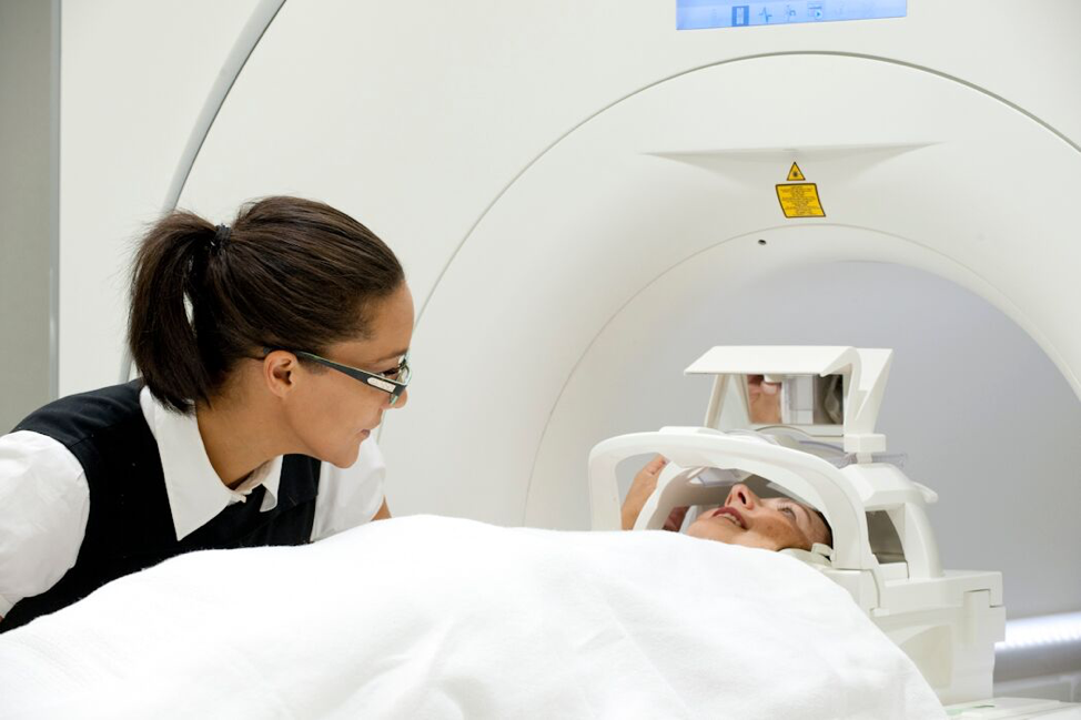
To provide this imaging information, a person will lie still in a MRI scanner (pictured below) and either look at pictures, listen to noises, watch a movie, or even play a game, depending on the type of information the researcher is interested in. These tasks can last anywhere from a few minutes to a couple hours, and all the while the MRI machine tracks blood flow in the brain and sends this information to a computer that creates images. Once the scan is complete, researchers or medical professionals use specialized data processing techniques that allow for meaningful interpretation of the images.
When proper safely procedures are followed, fMRI is non-invasive and low risk, which makes it a great (albeit expensive) research tool for use with both adults and children. In the first 5 years of life, the brain develops at an exponential rate, undergoing multiple developmental shifts. To better understand how the brain develops and the early experiences that lead to healthy development, fMRI provides an excellent lens into the growing mind. However, researchers usually implement special routines with children, as the look and sounds of a scanner can feel a bit novel, and staying still can be a challenge for some young children. For instance, researchers might need to take lots of time and incremental steps to let children warm up to the idea of a brain scan. With high quality warm-ups, about 80-90% of children can complete an MRI scan (3).
How do magnets make pictures?
Your brain is composed of many different nerve cells called neurons. In response to external sensory input, such as sight or touch, nerve cells are activated, which cause a small electrical signal that hops from neuron to neuron until it reaches a particular part of the brain. Think of it like a neural relay race, with each neuron passing the baton to the next until the signal reaches the part of the brain that is specialized to best interpret it.
To give us a relative idea of both when and where neural activity is occurring, fMRI measures these electrical signals in a creative way. For our relay runners (i.e., neurons) to continue their race, they need fuel. That fuel is delivered through blood cells, which supply neurons with essential nutrients like oxygen and glucose (broken-down sugar). Whenever bundles of neurons are activated in a specific area, blood flow increases to that area to restore those nutrients. Conveniently, blood cells have magnetic properties that translate into a blood-oxygen-level-dependent (BOLD) signal. During a scan, fMRI captures BOLD signals and creates a picture of when and where the brain is most active. fMRI can provide information about brain activation both in response to specific sensory inputs or when the brain is at rest.
Task-Based fMRI
Task-based fMRI is used to study neural signals that occur in response to an external event or sensory input (e.g., sounds, pictures, or playing a game). For example, a researcher might use task-based fMRI to investigate which areas of the brain are responsible for differentiating between speech sounds. To do this, the researcher might have participants undergo fMRI while they listen to two recordings – one of words being spoken in a person’s native language and another of words spoken in their non-native language. Then, by comparing the fMRI images generated during the two recordings, the researcher can detect differences in the activation of brain regions. These images could help the researcher discover if the brain works in a different way when it hears a non-native language, and if there are different areas of the brain that are specialized for interpreting each type of sound.
Resting state fMRI
Even when your brain isn’t receiving specific input (like the speech sounds in the task-based fMRI example), your neurons are still at work! The other type of fMRI, resting state fMRI, can provide information about the pathways that neurons are using in a state of relative rest. Neurons continuously reorganize themselves along frequently-activated pathways to run more efficient relays. To store memories, neurons that are activated during an event will make slight organizational changes, which will allow stored information to be accessed in the future. This is why repetition is helpful when learning a new skill – the more a pathway is activated, the more efficient neurons can be at running their race – such as by helping you remember a fact or carry out a task. Neurons are also constantly communicating about basic bodily functions, like heart rate, breathing, and hunger. Resting state fMRI provides a glance into the basic organization and wiring of brain networks, which lays the groundwork for how different inputs are processed.
Researchers can also use resting state fMRI to understand relative differences in brain networks for people who struggle with a specific cognitive ability. As an example, persons with attention-deficit/hyperactivity disorder (ADHD) usually have difficulties paying attention and controlling impulsive behavior. One study using resting state fMRI found that compared to adults without ADHD, adults with ADHD have different patterns of connectivity in brain networks responsible for both attention and reward (4). These types of findings are important because they often have practical implications for clinical practice. In the case of ADHD, this research suggests that therapies targeting both attention and motivation will be most beneficial.
A different snapshot
Just like any other research method, fMRI offers only one specific snapshot into the inner workings of the mind. fMRI is most useful when combined with other information, like data from observations and surveys, in order to paint a fuller picture. The brain doesn’t exist within a closed loop, but constantly interacts with behavior and the environment. fMRI allows researchers to observe these interactions in real time, and ultimately learn how the brain manages these interactions.
References
(1) Marinsek, N. (2017, December 18, 2017). 30 years of trends in the MRI and fMRI literatures. [Blog post]. Retrieved from: https://nikkimarinsek.com/blog/fmri-bursts
(2) Pfaff, D. W., & Volkow, N. D. (Eds.). (2016). Neuroscience in the 21st century: From basic to clinical. Springer.
(3) de Bie, H. M., Boersma, M., Wattjes, M. P., Adriaanse, S., Vermeulen, R. J., Oostrom, K. J., … & Delemarre-Van de Waal, H. A. (2010). Preparing children with a mock scanner training protocol results in high quality structural and functional MRI scans. European Journal of Pediatrics, 169(9), 1079-1085.
(4) Tomasi, D., & Volkow, N. D. (2012). Abnormal functional connectivity in children with attention-deficit/hyperactivity disorder. Biological Psychiatry, 71(5), 443-450.
Featured Images

[…] information about brain activation during a particular experience (Read more about this technique here). In this study, the researchers read the children stories while in the fMRI scanner to examine how […]
LikeLike
[…] activity, as more active tissues require more oxygen (4) (see previous post for more information https://cogbites.org/2019/07/22/what-is-fmri/). Increased startle response in PTSD patients was linked to higher LC activity in MRI scans, […]
LikeLike