
Reference: de Lange, A. G., Barth, C., Kaufmann, T., Maximov, I. I., Meer, D., Agartz, I., & Westlye, L. T. (2020). Women’s brain aging: Effects of sex‐hormone exposure, pregnancies, and genetic risk for Alzheimer’s disease. Human Brain Mapping, 41(18), 5141–5150.
Every day, millions of women are prescribed sex hormones. Although these drugs may have short-term benefits, the long-term impact on women’s health, especially their brain health, are not well known.
Female Sex Hormones
Sex hormones are either endogenous or exogenous; ENDOgenous hormones are produced inside the body and EXOgenous hormones are synthesized outside of the body (i.e., human made). Estrogens and progesterone are the two types of female endogenous hormones. They are created in the female ovaries and each naturally fluctuate during the course of a woman’s life. Exogenous hormones are ingested (e.g., orally) and interrupt these natural fluctuations by either increasing or decreasing endogenous hormone production.
Oral Contraceptives and Hormone Replacement Therapy
Oral contraception (abbreviated OC; an example is the birth control pill) and hormone replacement therapy (HRT) are the two most common exogenous hormonal drugs taken by women. OCs have been available for over 60 years (1) and are used by women around the world (2). Other than the obvious immediate benefits of OCs, such as providing a form of birth control, OCs have been demonstrated to enhance performance on verbal memory tasks (3). HRTs have also been available for decades and are generally prescribed to alleviate negative menopausal symptoms, but unlike OCs, HRTs have received more attention in the research world because of emerging evidence linking HRT use and reduced Alzheimer’s disease risk (4). However, the long-term effects of both OCs and HRTs on women’s health are understudied.
Current research on women’s brain health and sex hormone exposure
Over the past decade, research on the effects of endogenous hormones on women’s brain health has gained some momentum. For example, studies have illustrated various effects that endogenous hormones have on a woman’s brain’s structure, function, and cognitive performance at different transitional periods of her life (5, 6). Specifically, during a woman’s reproductive years, higher levels of endogenous hormones are associated with larger brain volumes (7, 8) and changes in hormone fluctuations are known to influence brain resiliency (9, 10). In addition, women who have given birth to a child have demonstrated a younger-looking brain compared to women who have not experienced a pregnancy (11).
Exogenous hormone research, on the other hand, is less concrete. HRTs have been shown to have some protective effects against neurodegenerative diseases such as Alzheimer’s and Parkinson’s disease (12, 13); but, there is growing research indicating that these effects are only evident if HRT is taken at a specific point in a woman’s life. The “critical period hypothesis” states that if HRT is initiated soon after menopause begins (i.e., within 5 years) then it can be protective against cognitive decline however, if HRT is taken later in menopause, then it could be detrimental to cognition (14). Furthermore, HRT-users 65 years and older have demonstrated greater brain shrinkage compared to non-users of the same age (15)
Are there long-term effects of sex hormone exposure?
To help bridge this gap in women’s brain research and sex hormone exposure, a recent study by de Lange and colleagues set out to investigate multiple associations between endogenous and exogenous sex hormones and their effect on normal brain aging. Data from more than 16,000 women between the ages of 40 to 70 years were obtained from the United Kingdom Biobank.
The paper included many different analyses but I will only focus on four in this blog post.
First, the women’s apparent brain age was calculated by subtracting their chronological (i.e., actual) age from their estimated brain age. Estimated brain age was calculated through the use of machine learning and functional magnetic resonance imaging (i.e., fMRI) measures such as brain structure thickness and volume. Therefore, if a 50-year-old woman’s brain demonstrated brain characteristics of a typical 70-year-old, then their apparent brain age would be higher than their chronological age.
Second, cumulative sex hormone (both exogenous and endogenous) exposure was estimated by an index of cumulative estrogen exposure (16), which was calculated using a metric that considered the age at first period, when menopause began, time since menopause, body mass index, and the duration of HRT use.
Third, exogenous hormone exposure was estimated by HRT and OC use.
Finally, to assess the “critical period hypothesis” of HRT, age at HRT initiation, both alone and in relation to age at menopause, and apparent brain aging were examined. Because de Lange and colleagues (11) previously assessed the relationship between women who had given birth to a child compared to women who had not, all analyses were adjusted for childbirth status.
Summary of Key Findings
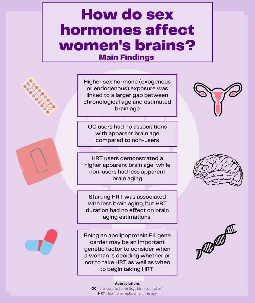
The study illustrated that increased sex hormone exposure was linked to higher apparent brain age which means that women with more cumulative hormone (endogenous and/or exogenous) exposure over the life span (indexed as age at first period, when menopause began, time since menopause, body mass index, and the duration of HRT use) had a larger gap between their chronological age and their apparent brain age.
For exogenous hormones, OC users compared to non-users had no association with apparent brain age. Thus, taking OCs did not have an association with women’s brain health when using apparent brain estimations as the method of measurement. On the other hand, HRT users demonstrated a higher apparent brain age in both premenopausal and menopausal women while non-users had less apparent brain aging. This suggests that HRT use may be detrimental to some women causing their brains to appear ‘older’ then their chronological age. But, earlier HRT initiation was positively associated with less brain aging. Therefore, women who started HRT earlier had a brain that appeared similar to what would be expected for the women’s chronological age compared to women who started HRT treatment later. HRT duration, on the other hand, had no effect on brain aging estimations.
Furthermore, to assess whether there was a particular association between HRT use in women with a genetic risk factor for Alzheimer’s disease, women with and without a specific genotype (apolipoprotein E4) were compared. It was shown that carriers (those who carried the gene) who began HRT before the onset of menopause demonstrated less evident brain aging compared to non-carriers (those who did not carry the gene). When looking at carriers only, those with high levels of endogenous estrogen demonstrated more apparent brain aging compared to carriers with low levels of endogenous estrogen during menopause. But, non-carriers with low levels of endogenous estrogens demonstrated more apparent brain aging compared to non-carriers with high levels of endogenous estrogen during menopause. This suggests that genetic factors may be important to consider for women’s brain health when deciding if and when to begin taking HRT..
So what can we take away from these results?
Broadly speaking, this research highlights how high levels of sex hormone (both endogenous and exogenous) exposure may have adverse effects on women’s brain aging. Consistent with the critical period hypothesis, earlier age at HRT initiation was associated with less evident brain aging; however this was only the case for women carriers of a specific gene. Neither HRT duration nor OC status appeared to have any effect on brain aging.
Future Directions
Future research should consider the many different types of OCs and HRTs (i.e., estrogens only or combination of estrogens and progesterone) and methods of ingestion (i.e., oral or transdermal), as these factors may have different effects on the brain. Moreover, it would be valuable to take into account that OC use is commonly taken in cycles (i.e., when a women is trying to avoid or promote pregnancy) because the fluctuations in hormones may have positive (or negative) effects to a woman’s brain health. Also, breastfeeding is known to reduce exposure to endogenous hormones, so analyzing the brains of women who breastfeed compared to those who don’t and following these women across time would provide a more concrete estimation of cumulative sex hormone exposure. Last, longitudinal studies with more detailed information on all of the variables included in the current research would further the understanding of how sex hormone exposure influences women’s brain health across their lifespan.
Conclusions
All in all, the impact of sex hormone exposure on women’s brain health requires further study, however the ground-breaking studies published over the past decade have built a solid foundation for future work and the goal of having a positive impact on women’s health appears attainable!
Additional References:
- Pletzer, B. A., & Kerschbaum, H. H. (2014). 50 years of hormonal contraception -”time to find out, what it does to our brain. Frontiers in Neuroscience, 8. https://doi.org/10.3389/fnins.2014.00256
- Petitti, D. B. (2003). Combination Estrogen–Progestin Oral Contraceptives. New England Journal of Medicine, 349(15), 1443–1450. https://doi.org/10.1056/NEJMcp030751
- Mordecai, K. L., Rubin, L. H., & Maki, P. M. (2008). Effects of menstrual cycle phase and oral contraceptive use on verbal memory. Hormones and Behavior, 54(2), 286–293. https://doi.org/10.1016/j.yhbeh.2008.03.006
- Yaffe, K., Haan, M., Byers, A., Tangen, C., & Kuller, L. (2000). Estrogen use, APOE, and cognitive decline: Evidence of gene–environment interaction. Neurology, 54(10), 1949–1954. https://doi.org/10.1212/WNL.54.10.1949
- Toffoletto, S., Lanzenberger, R., Gingnell, M., Sundström-Poromaa, I., & Comasco, E. (2014). Emotional and cognitive functional imaging of estrogen and progesterone effects in the female human brain: A systematic review. Psychoneuroendocrinology, 50, 28–52. https://doi.org/10.1016/j.psyneuen.2014.07.025
- Rehbein, E., Hornung, J., Sundström Poromaa, I., & Derntl, B. (2020). Shaping of the female human brain by sex hormones – a review. Neuroendocrinology. https://doi.org/10.1159/000507083
- Barth, C., Steele, C. J., Mueller, K., Rekkas, V. P., Arélin, K., Pampel, A., Burmann, I., Kratzsch, J., Villringer, A., & Sacher, J. (2016). In-vivo Dynamics of the Human Hippocampus across the Menstrual Cycle. Scientific Reports, 6(1), 32833. https://doi.org/10.1038/srep32833
- Lisofsky, N., Mårtensson, J., Eckert, A., Lindenberger, U., Gallinat, J., & Kühn, S. (2015). Hippocampal volume and functional connectivity changes during the female menstrual cycle. NeuroImage, 118, 154–162. https://doi.org/10.1016/j.neuroimage.2015.06.012
- Galea, L. A. M., Leuner, B., & Slattery, D. A. (2014). Hippocampal Plasticity during the Peripartum Period: Influence of Sex Steroids, Stress and Ageing. Journal of Neuroendocrinology, 26(10), 641–648. https://doi.org/10.1111/jne.12177
- Simerly, R. B. (2002). Wired for Reproduction: Organization and Development of Sexually Dimorphic Circuits in the Mammalian Forebrain. Annual Review of Neuroscience, 25(1), 507–536. https://doi.org/10.1146/annurev.neuro.25.112701.142745
- de Lange, A.-M. G., Kaufmann, T., van der Meer, D., Maglanoc, L. A., Alnæs, D., Moberget, T., Douaud, G., Andreassen, O. A., & Westlye, L. T. (2019). Population-based neuroimaging reveals traces of childbirth in the maternal brain. Proceedings of the National Academy of Sciences, 116(44), 22341–22346. https://doi.org/10.1073/pnas.1910666116
- Erickson, K. I., Colcombe, S. J., Raz, N., Korol, D. L., Scalf, P., Webb, A., Cohen, N. J., McAuley, E., & Kramer, A. F. (2005). Selective sparing of brain tissue in postmenopausal women receiving hormone replacement therapy. Neurobiology of Aging, 26(8), 1205–1213. https://doi.org/10.1016/j.neurobiolaging.2004.11.009
- Ha, D. M., Xu, J., & Janowsky, J. S. (2007). Preliminary evidence that long-term estrogen use reduces white matter loss in aging. Neurobiology of Aging, 28(12), 1936–1940. https://doi.org/10.1016/j.neurobiolaging.2006.08.007
- MacLennan, A. H., Henderson, V. W., Paine, B. J., Mathias, J., Ramsay, E. N., Ryan, P., Stocks, N. P., & Taylor, A. W. (2006). Hormone therapy, timing of initiation, and cognition in women aged older than 60 years: The REMEMBER pilot study. Menopause, 13(1), 28–36. https://doi.org/10.1097/01.gme.0000191204.38664.61
- Resnick, S. M., Espeland, M. A., Jaramillo, S. A., Hirsch, C., Stefanick, M. L., Murray, A. M., Ockene, J., & Davatzikos, C. (2009). Postmenopausal hormone therapy and regional brain volumes: The WHIMS-MRI Study. Neurology, 72(2), 135–142. https://doi.org/10.1212/01.wnl.0000339037.76336.cf
- Smith, C. A., McCleary, C. A., Murdock, G. A., Wilshire, T. W., Buckwalter, D. K., Bretsky, P., Marmol, L., Gorsuch, R. L., & Buckwalter, J. G. (1999). Lifelong Estrogen Exposure and Cognitive Performance in Elderly Women. Brain and Cognition, 39(3), 203–218. https://doi.org/10.1006/brcg.1999.1078

[…] After a year of travel, Alex continued her academic journey by completing a Master of Arts in Psychology from the University of Toronto. Her master’s thesis explored risk factors involved in the development of mild cognitive impairment and Alzheimer’s disease. Currently, as a Ph.D. student, Alex is working on multiple projects including the investigation of functional and structural changes that occur in neurodegeneration and healthy aging as well as looking at sex differences in an at-risk Alzheimer’s disease population. Her most recent post on cogbites was about how sex hormones affect women’s brains. […]
LikeLike