
Reference: Dong, W. Y., Zhu, X., Tang, H. D., Huang, J. Y., Zhu, M. Y., Cheng, P. K., … & Zhang, Z. (2023). Brain regulation of gastric dysfunction induced by stress. Nature Metabolism, 5(9), 1494-1505.
What’s the Societal Relevance?
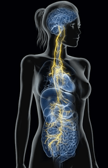
You’ve probably noticed while walking through your local supermarket the newest marketing rave: food and beverages promoting “gut health”. With rows and rows of brightly colored kombuchas, prebiotic soda alternatives, and supplements lining the shelves, you may be wondering whether there is science behind this craze. You may also be curious how adverts suggest improving our “gut health” may be beneficial for more than just weight loss, but also make us generally feel better. For decades the stomach was considered separate from the brain, with gastric research focusing mostly on pathology directly impacting the stomach (e.g., bacterial infections and stomach cancers) as opposed to any neuronal influences. With over 100 million nerve cells lining the gastrointestinal (G.I.) tract, there is rapid communication between our central nervous system (CNS, which consists of the brain and spinal cord) and our enteric nervous system (ENS, which consists of neurons within the gastrointestinal tract). It has long been thought that anxiety and depressive disorders contributed to G.I. disruption, but recent findings show it may be the other way around, with our stomach influencing our brain’s functions that ultimately impact mood, cognition, and behavior. Stress has many gastric and neuronal effects. Researchers Wan-Ying Dong and colleagues recently uncovered the specific pathway the brain connects to the stomach and how stress can change that connection.
The Overlap of Stress and Gastric Disorders
You may be familiar with the phrase, “rest and digest”, commonly paired with the phrase, “fight or flight.” These phrases are our bodies’ common reaction to low or high stress respectively. The Vagus nerve is a massive blood transporter, traveling all throughout the body. The Vagus nerve is primarily responsible for inducing a relaxed state where our bodies focus on getting nutrients and energy from food to prepare for the next time we need to do something physically demanding (1). Acetylcholine is the exact molecule that released by the Vagus nerve causing this effect, which I will refer to as the “rest and digest signaler”. Acetylcholine opposes adrenaline, the fight or flight signaler, released by our adrenal glands to prepare for strenuous physical activity. These signalers have opposing effects on different organs. Stress activates the fight or flight signaler, which eventually results in diverting resources from digestion towards movement. Fight or flight signaling betters our ability to move by increasing heart rate, increasing lung capacity, and increasing sugar release into the bloodstream while stomach activity is reduced and bowel movements are increased.
Many gastric disorders reflect the effects of stress on the digestive system. For example, functional dyspepsia is a common stomach disorder where food is digested slower than normal, and irritable bowel syndrome (IBS) is often characterized by increased bowel movements. About 12% of the world has IBS, with anxiety and depression occurring in almost 40% of those patients (2). Studies have shown that IBS patients have increased sensitivity to stress-inducing bowel movements. For example, when IBS patients are given a stress hormone injection or hear a psychologically stressful phrase (e.g., “you’re late”), they are more likely to have to use the restroom soon after, whereas this does not happen to healthy participants (2). This outcome suggests that people with IBS are more sensitive to the digestive impacts of stress, but where and how this stress sensitivity arises remains unclear.
Researchers are currently trying to better understand the role of prolonged stress in gastric dysfunction. Gastric dysfunction can be induced by chronic stress in animal models, but exactly how the brain is involved in this process had previously been unclear. The Zhang laboratory used modern neuroscience tools to map out pathways that link the brain and gut after persistent stress.
Connecting the Brain to Our Gut
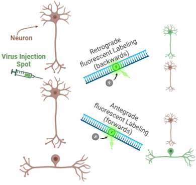
Researchers used viral tract tracing, a technique that uses different viruses traveling with or against a neuron’s electric signaling path, a neuron being a tiny nerve cell in the brain that receives and sends information. The researchers cause specific neurons and their root-like endings to glow with fluorescence in order to image their location and interaction in the gut (see the figure below). In viewing the fluorescence, researchers discovered a brain region called the dorsal raphe nucleus filled with neurons containing serotonin, connects to the Vagus nerve that signals directly to the stomach. Serotonin is often known as a mood stabilizer or the “happy hormone” and is targeted in the treatment of depression. Serotonin is also actively involved in increasing digestion, controlling satiety (the feeling when you are full from a meal), and nausea (4). This pathway connecting our brain’s supply of the happy hormones to the signalers for resting and digesting may be of interest in the treatment of IBS, especially for patients who also struggle with mood disorders.
What Happens After Chronic Stress?
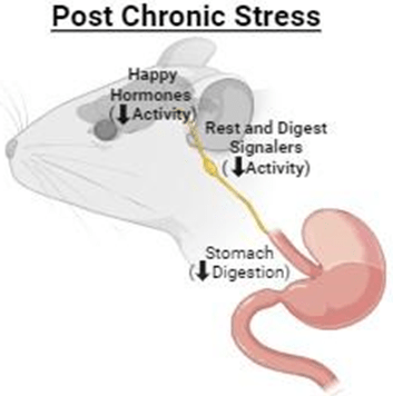
Viral tract tracing revealed that the brain and the gut are linked. The next step for Zhang and colleagues was to understand how a chronically stressful state can change the activity of the brain and its signaling to the gut. Researchers combined viral tract tracing with techniques used to measure neuronal activity to understand what changes occur in the brain sources for happy hormones and rest and digest signaling after chronic stress. They used an animal model (mice) of chronic stress to do this. For two weeks, animal cages were placed on a slight tilt at all times, and for one hour each day they were placed in a puddle of water for 45 minutes followed by having their tails pinched by a paper clamp for 15 minutes. These animals had reduced activity in the brain regions containing happy hormones (serotonin) and rest and digest signalers (acetylcholine) when compared to animals that were not stressed. Stomach activity was also reduced in the stressed animals, causing food to sit in the stomach for longer, and therefore the animals ate less.
Optogenetics was a tool used to control brain activity in this study. Optogenetics genetically inserts an opening activated by a certain color of light (i.e., blue) that increases brain activity. Stimulating the happy hormone source that connected to the rest and digest signalers to the stomach with blue light improved digestion in chronically stressed animals. Scientists also used chemogenetics, another genetic tool to alter brain activity. Chemogenetics uses a chemical rather than light, the chemical being a drug designed by scientists that only they can activate. Researchers injected their special chemical into the animal to increase the activity of the same happy hormone to rest and digest signalers once again, also improving digestion. Both ways of increasing the activity of this specific area of happy hormones improved digestion in the chronically stressed group.
What About Acute Stress?
An acute stressor implemented for only one hour, as opposed to the two-week duration of the chronic stressors, had the opposite effects of chronic stress. That is, acute stress increased the activity of happy hormone and rest and digest signal sources, which allowed for food to move through the digestive tract faster and for the animals to eat more. This study exemplified how the duration and type of stress can have opposing impacts on gastric function, which is important for understanding gastrointestinal conditions such as IBS.
Future Directions
There are still many questions left for researchers to investigate regarding how our brain and gut communicate. About 95% of the body’s serotonin (or happy hormone) is produced in the gut, but little is known about how the serotonin in our gut communicates with our brain (4). Commonly used antidepressants that target the serotonergic system have been shown to disrupt gut function, suggesting the way these drugs act in the brain versus the gut is different and should be investigated further so that people taking these drugs are aware of the full effects. Investigating whether gut-health promoting supplements help alleviate negative gastric effects would be beneficial to improve the overall quality of health for these patients. Understanding the role of serotonin in the gut and the brain is especially relevant for the many IBS patients who also have depression and other stress-sensitive psychiatric disorders treated with serotonin-targeting drugs. Overall, understanding how our brain and gut communicate is important for improving future outcomes for both gastric and psychiatric patient care.
Additional References:
(1) Dolphin, H., Dukelow, T., Finucane, C., Commins, S., McElwaine, P., & Kennelly, S. P. (2022). “The Wandering Nerve Linking Heart and Mind” – The Complementary Role of Transcutaneous Vagus Nerve Stimulation in Modulating Neuro-Cardiovascular and Cognitive Performance. Frontiers in Neuroscience. 16(16), 897303. https://dio.org/10.3389/fnins.2022.897303.
(2) Mertz, H. Stress and the Gut. UNC Center for Functional GI and Motility Disorders.
(3) Ormsbee, H. S., 3rd, & Fondacaro, J. D. (1985). Action of serotonin on the gastrointestinal tract. Proceedings of the Society for Experimental Biology and Medicine. Society for Experimental Biology and Medicine (New York, N.Y.), 178(3), 333–338. https://doi.org/10.3181/00379727-178-42016
(4) Terry N. & Margolis, K. G. (2017). Serotonergic Mechanisms Regulating the GI Tract: Experimental Evidence and Therapeutic Relevance. Handbook of Experimental Pharmacology. 239, 319-342. https://dio.org/10.1007/164.2016.103.
