
Occasionally forgetting a word or stumbling over a sentence can be a normal part of life, particularly as we age. But for people with primary progressive aphasia (PPA), difficulties with language and communication impact more than just a single conversation or moment in time.
The life-altering reality of the disorder has been illustrated through the diagnosis of notable celebrities Bruce Willis and Wendy Williams, who have stepped out of the spotlight in recent years due to increasing difficulties with language and communication. These communication challenges are broadly captured under the term “aphasia”, but for those with PPA, such difficulties become more common and severe over time. A person with PPA may frequently forget words, struggle to remember the name of a familiar person or object, or have trouble following a conversation. These complications make basic daily activities challenging, and can greatly impact the lives of those diagnosed and their loved ones.
Despite the media spotlight from these prominent cases, PPA is still relatively unknown by the vast majority, and misconceptions about the disorder are common. As dementia becomes more prevalent in an aging population, it is more important than ever to understand the conditions that affect those diagnosed and their loved ones.
Understanding and Diagnosing PPA
The term “primary progressive aphasia”, or PPA, refers to a type of dementia that primarily affects speech and language (Ruksenaite et al., 2021). Primary refers to the fact that difficulties with language are the earliest and most prominent symptom, while progressive means that the symptoms worsen over time, and aphasia refers to difficulty using or understanding language.
The disorder develops when damage related to cell death occurs in the brain, also known as neurodegeneration. This damage is caused by neurodegenerative diseases, particularly Alzheimer’s disease and frontotemporal lobar degeneration, which can also progress to frontotemporal dementia. Although PPA is often related to these diseases, it is a separate clinical syndrome with distinct symptoms. PPA is typically diagnosed earlier than other forms of dementia, appearing in most cases by age 65 (Ruksenaite et al., 2021). In patients who have been diagnosed, loss of brain tissue and function occurs primarily in language-dominant regions, which is why PPA primarily affects speech and language early in the disease course.
Although PPA is a distinct condition from other types of dementia, symptoms may still vary from patient to patient. Three variants of PPA are currently recognized, each with its own clinical presentation (Table 1).
Nonfluent/agrammatic variant PPA is typically recognizable in affected individuals who speak at a significantly slower rate than conversational pace. Patients may make basic grammar or syntax mistakes, a disorder known as agrammaticism. While patients diagnosed with semantic variant PPA may also display difficulty with basic communication, patients with this variant typically present with a different condition known as anomia, meaning they have difficulty naming familiar people and objects. Individuals living with the semantic variant of PPA often report that they “know” an object, but cannot “say” what it is (Mesulam et al., 2014).
The final subtype is known as logopenic variant PPA. Affected individuals have difficulty recalling words during spontaneous conversation, and struggle with repeating sentences spoken aloud. They may also have trouble with basic reading, spelling, and sentence comprehension, sometimes leading to a diagnosis of dyslexia or other reading disorders.
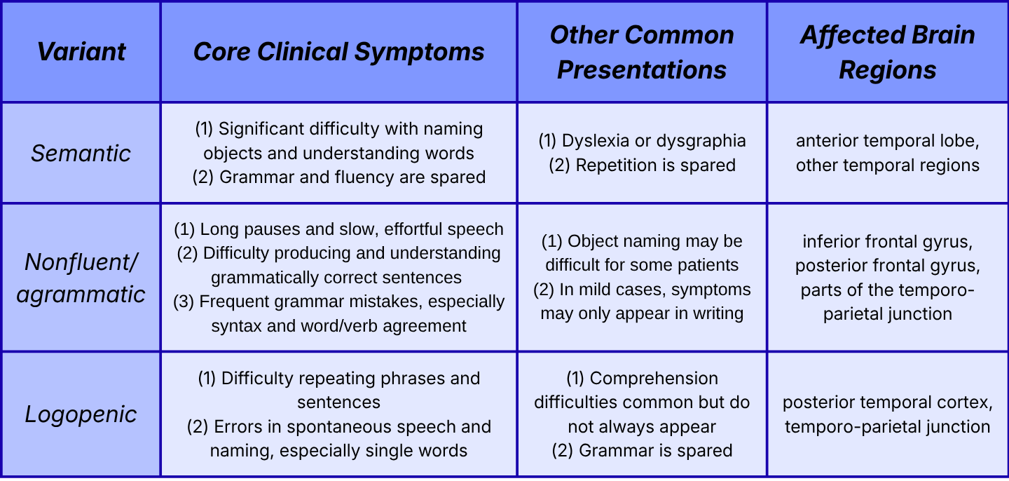
While distinct language difficulties are unique hallmarks of PPA, it is common for other symptoms to appear over time. Severe cognitive decline, motor deficits, and behavioral disturbances usually associated with the behavioral variant of frontotemporal dementia may present as the disease progresses (Ulugut et al., 2022). The appearance of other symptoms related to different forms of dementia complicates accurate identification and diagnosis of PPA, and presents a unique challenge for doctors treating affected individuals.
Why is PPA Difficult to Diagnose?
Difficulties with word finding, deficits in verbal short-term memory, and general decline in communication skills are just a few symptoms commonly noted in many presentations of dementia. Because of the significant overlap in speech and language symptoms for PPA and other dementias, physicians must rely on distinct criteria to differentiate between disorders.
In the case of PPA, the challenge of identification and diagnosis stems from the complexity and variability of the disease. Patients with PPA, especially those who present with mixed symptoms, are frequently misdiagnosed with psychiatric disorders, Alzheimer’s disease, or other neurodegenerative conditions (Clarke & Bonadarpour, 2019; Mulder-Heijstra et al., 2022).
The Problem of Misdiagnosis
Misdiagnosis presents a significant problem for individuals affected by PPA. Achieving an early, accurate diagnosis is crucial for life planning and access to critical support and services (Ruksenaite et al., 2021). Incorporating speech therapies as early as possible, such as word retrieval exercises, script therapy, and compensatory strategies, can help improve language abilities in patients diagnosed with PPA (Machado et al., 2021; Volkmer et al., 2020; Wauters et al., 2024).
For many of these patients, therapeutic support and services translate to substantial improvements in everyday living, leading to better communication and increasing quality of life. Still, the complexity of the disorder means that physicians cannot solely depend on language assessments. Early, accurate diagnosis of PPA relies on more detailed tools.
What the Brain Can Tell Us About PPA
The introduction of neurobiology to the management of neurodegenerative disorders has fundamentally shifted the way we identify, assess, and treat PPA (Curtis et al., 2025). When clinical diagnosis is unable to provide enough detail to make an accurate diagnosis, doctors can now consult imaging techniques which have been shown to help distinguish among PPA variants.
For most variants of PPA, symptoms can be related to frontotemporal degeneration, which refers to brain damage found in the frontal and temporal lobes that slowly worsens over time. Using modern imaging methods, this damage can now be observed as the condition progresses. Such methods include magnetic resonance imaging (MRI), which provides information about brain structure and volume, and F-fluorodeoxyglucose positron emission tomography (FDG-PET), which detects underlying abnormalities by tracking how the brain uses glucose (Giannakis et al., 2026; Ruksenaite et al., 2021). Both MRI and FDG-PET have been used to understand how symptoms of PPA are associated with damage to specific areas of the brain.
Connecting Symptoms with Neurobiology
Research has found that nonfluent/agrammatic PPA is associated with different areas of the brain than the semantic variant, a crucial step forward in understanding how different symptoms may be related to areas of damage. The semantic variant of PPA is primarily associated with anterior temporal lobe atrophy, which means that the brain tissue in this region shrinks or dies off, causing a breakdown in brain function (Figure 1). The nonfluent/agrammatic variant is linked to frontal regions, such as the inferior frontal gyrus, as well as some temporal and parietal areas. Atrophy in the temporo-parietal junction, or the area between the temporal and parietal lobes, is typically associated with the logopenic variant (Gorno-Tempini et al., 2011; Mesulam et al., 2014; Ruksenaite et al., 2021). Most importantly, these associations provide a pattern for physicians to look for when examining a patient’s brain.
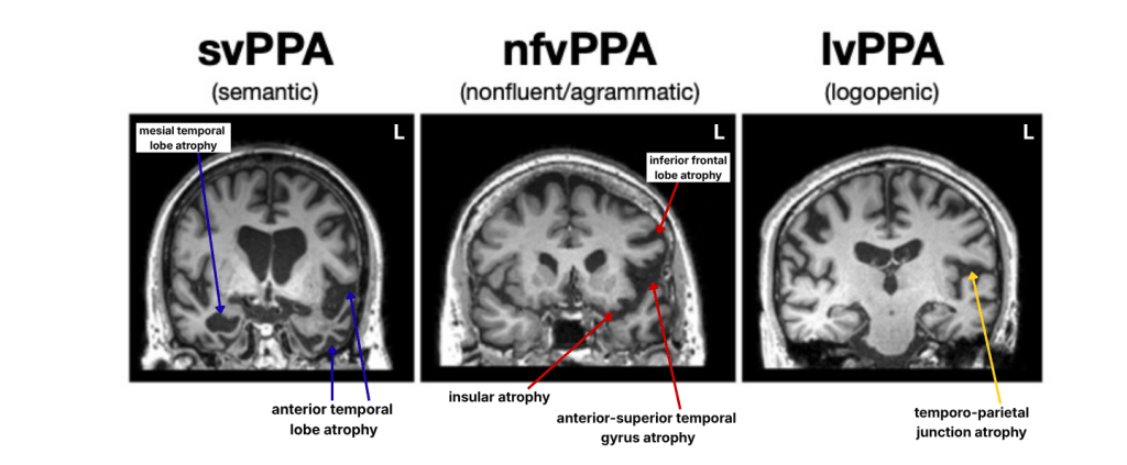
Imaging methods such as MRI can also link specific symptoms to damaged areas. Researchers have used MRI to uncover a new “information superhighway” which connects key language regions. This white matter pathway, known as the frontal aslant tract, has been linked to deficits in verbal fluency related to PPA (Catani et al., 2013). Studies have since used MRI to investigate specific areas of damage in relation to PPA symptoms (Behn et al., 2025), and even uncover new potential subtypes of PPA based on anatomy (Taylor et al., 2025). Imaging alone is not a perfect solution, but it is another instrument in a physician’s toolkit which can improve diagnostic accuracy and prevent treatment delays.
The final step in confirming a diagnosis of a neurodegenerative brain disease is analyzing the pathology, or looking at the actual brain tissue under a microscope to confirm cell death caused by Alzheimer’s disease or frontotemporal lobar degeneration. This step is the most invasive, and many pathophysiological features can only be investigated post-mortem since the brain tissue needed to diagnose the disease cannot be removed while the patient is alive. Thus, the question remains: what can be done to improve quality of life for patients while treatment is still an option?
Treating a Disease with No Cure
Beyond speech and language therapy, scientists are beginning to uncover ways of treating PPA using neurobiologically-informed methods. Non-invasive brain stimulation techniques, such as transcranial direct current stimulation and transcranial magnetic stimulation, send weak electrical currents into the brain to excite or inhibit brain activity in certain regions. These methods have shown promise in alleviating symptoms of PPA (Cotelli et al., 2020; Nissim et al., 2020), though such research is still in the early stages. Importantly, promising new techniques are being discovered every day to improve the lives of those affected by PPA and other neurodegenerative disorders.
PPA is a life-altering disease, not just for the individual diagnosed, but for friends, family members, and caregivers. Early evaluation and treatment are crucial for affected individuals to access speech-language therapy, communication tools, support groups, and educational resources, which can help loved ones and caregivers learn how to support someone living with PPA.
That support often means adapting how one communicates to aid in understanding and avoid frustration. Basic tips and strategies for communicating with an individual affected by PPA include: using direct language, keeping sentences short, breaking down directions into small steps, using gestures and pictures when applicable, and exercising patience by allowing the person time to communicate. To learn more about PPA and how to support a loved one affected by the condition, visit the following resources:
- American Speech-Language-Hearing Association: A simple breakdown of primary progressive aphasia.
- The Mesulam Institute for Cognitive Neurology & Alzheimer’s Disease: Information on the care, support, and treatment of PPA, as well as aphasia-friendly resources and guides.
- The Association for Frontotemporal Degeneration: Learn more about FTD/PPA and related diseases, find support, and get involved with research.
References:
(1) Behn, J. Q., Barbieri, E., Mesulam, M. M., & Bonakdarpour, B. (2025). Anterior temporal lobe, word comprehension, and physiology of atrophy in semantic primary progressive aphasia. Neurocase, 31(6), 260–267. https://doi.org/10.1080/13554794.2025.2587123
(2) Catani, M., Mesulam, M. M., Jakobsen, E., Malik, F., Martersteck, A., Wieneke, C., Thompson, C. K., Thiebaut de Schotten, M., Dell’Acqua, F., Weintraub, S., & Rogalski, E. (2013). A novel frontal pathway underlies verbal fluency in primary progressive aphasia. Brain, 136(Pt 8), 2619–2628. https://doi.org/10.1093/brain/awt163
(3) Clarke, E. C., & Bonakdarpour, B. (2019). Challenges in diagnosis of primary progressive aphasia. BMJ Case Reports, 12(11), e230630. https://doi.org/10.1136/bcr-2019-230630
(4) Cotelli, M., Manenti, R., Ferrari, C., Gobbi, E., Macis, A., & Cappa, S. F. (2020). Effectiveness of language training and non-invasive brain stimulation on oral and written naming performance in primary progressive aphasia: A meta-analysis and systematic review. Neuroscience and Biobehavioral Reviews, 108, 498–525. https://doi.org/10.1016/j.neubiorev.2019.12.003
(5) Curtis, M., Alfano, A. R., Murphy, D., Pruden, S. M., Bayat, M., Graziano, P. A., & Dick, A. S. (2025). Neurobiological contributions to speech and language interventions: Applications to developmental populations. Developmental Neuropsychology, 50(3-8), 185–214. https://doi.org/10.1080/87565641.2025.2581699
(6) Giannakis, A., Anyfantis, E., Litsou, E., Sioka, C., & Konitsiotis, S. (2026). FDG-PET in the evaluation of primary progressive aphasia: A narrative review. Medicina, 62(5), 800. https://doi.org/10.3390/medicina62050800
(7) Gorno-Tempini, M. L., Hillis, A. E., Weintraub, S., Kertesz, A., Mendez, M., Cappa, S. F., Ogar, J. M., Rohrer, J. D., Black, S., Boeve, B. F., Manes, F., Dronkers, N. F., Vandenberghe, R., Rascovsky, K., Patterson, K., Miller, B. L., Knopman, D. S., Hodges, J. R., Mesulam, M. M., & Grossman, M. (2011). Classification of primary progressive aphasia and its variants. Neurology, 76(11), 1006–1014. https://doi.org/10.1212/WNL.0b013e31821103e6
(8) Machado, T. H., Carthery-Goulart, M. T., Campanha, A. C., & Caramelli, P. (2021). Cognitive intervention strategies directed to speech and language deficits in primary progressive aphasia: Practice-based evidence from 18 cases. Brain Sciences, 11(10), 1268. https://doi.org/10.3390/brainsci11101268
(9) Mesulam, M. M., Rogalski, E. J., Wieneke, C., Hurley, R. S., Geula, C., Bigio, E. H., Thompson, C. K., & Weintraub, S. (2014). Primary progressive aphasia and the evolving neurology of the language network. Nature Reviews Neurology, 10(10), 554–569. https://doi.org/10.1038/nrneurol.2014.159
(10) Mouton, A., Plonka, A., Fabre, R., Tran, T. M., Robert, P., Macoir, J., Manera, V., & Gros, A. (2022). The course of primary progressive aphasia diagnosis: a cross-sectional study. Alzheimer’s Research & Therapy, 14(1), 64. https://doi.org/10.1186/s13195-022-01007-6
(11) Mulder-Heijstra, M. M. P., Jokel, R. R., Chertkow, H. H., Conn, D. D. K., & Mah, L. L. (2022). Primary progressive aphasia presenting with neuropsychiatric symptoms. Journal of Geriatric Psychiatry and Neurology, 35(4), 574–579. https://doi.org/10.1177/08919887211036189
(12) Nissim, N. R., Moberg, P. J., & Hamilton, R. H. (2020). Efficacy of noninvasive brain stimulation (tDCS or TMS) paired with language therapy in the treatment of primary progressive aphasia: An exploratory meta-analysis. Brain Sciences, 10(9), 597. https://doi.org/10.3390/brainsci10090597
(13) Ruksenaite, J., Volkmer, A., Jiang, J., Johnson, J. C., Marshall, C. R., Warren, J. D., & Hardy, C. J. (2021). Primary progressive aphasia: Toward a pathophysiological synthesis. Current Neurology and Neuroscience Reports, 21(3), 7. https://doi.org/10.1007/s11910-021-01097-z
(14) Taylor, B., Bocchetta, M., Shand, C., Todd, E. G., Chokesuwattanaskul, A., Crutch, S. J., Warren, J. D., Rohrer, J. D., Hardy, C. J. D., & Oxtoby, N. P. (2025). Data-driven neuroanatomical subtypes of primary progressive aphasia. Brain, 148(3), 955–968. https://doi.org/10.1093/brain/awae314
(15) Ulugut, H., Stek, S., Wagemans, L. E. E., Jutten, R. J., Keulen, M. A., Bouwman, F. H., Prins, N. D., Lemstra, A. W., Krudop, W., Teunissen, C. E., Van Berckel, B. N. M., Ossenkoppele, R., Barkhof, F., Van Der Flier, W. M., Scheltens, P., & Pijnenburg, Y. A. L. (2022). The natural history of primary progressive aphasia: Beyond aphasia. Journal of Neurology, 269(3), 1375–1385. https://doi.org/10.1007/s00415-021-10689-1
(16) Volkmer, A., Rogalski, E., Henry, M., Taylor-Rubin, C., Ruggero, L., Khayum, R., Kindell, J., Gorno-Tempini, M. L., Warren, J. D., & Rohrer, J. D. (2020). Speech and language therapy approaches to managing primary progressive aphasia. Practical Neurology, 20(2), 154–161. https://doi.org/10.1136/practneurol-2018-001921
(17) Wauters, L. D., Croot, K., Dial, H. R., Duffy, J. R., Grasso, S. M., Kim, E., Schaffer Mendez, K., Ballard, K. J., Clark, H. M., Kohley, L., Murray, L. L., Rogalski, E. J., Figeys, M., Milman, L., & Henry, M. L. (2024). Behavioral treatment for speech and language in primary progressive aphasia and primary progressive apraxia of speech: A systematic review. Neuropsychology Review, 34(3), 882–923. https://doi.org/10.1007/s11065-023-09607-1

